Core Equipments of IMIL
OlympusFluovieww Confocall LASER Scanning Microscope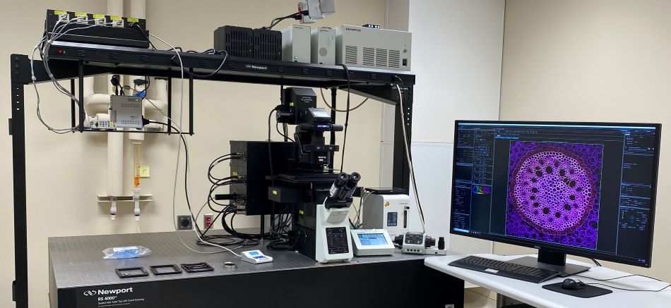
- Olympus IX833 fully motorized inverted microscope with ZDCC LASER-based autofocuss
- Resonant scanner for high-speed acquisition and galvanometer scanner for high definition
- Automated microscope stage
- 4 High-Sensitivity GaAsP detectors
- Tokaii hit stage-top live-cell incubation chamber
- Transmitted detector for Brightfieldd C image acquisition
- Analog and digital in-out box for synchronization with external devices
Available Laser Lines (nm): 405-445-488-514-561-640
Objectives
| Objective (UV corrected) | Mag. | NA | Immersion | WD (μm) | DICC | Color ID |
|---|---|---|---|---|---|---|
| PlanApo | 2x | 0.08 | dry | 6200 | No | white |
| U Plan SApoo | 10xx | 0.4 | dry | 3000 | Yes* | yellow |
| U Plan SApoo | 20xx | 0.75 | dry | 600 | Yes | green |
| U Plan SApoo N W/CC | 30xx | 1.05 | silicone oil | 800 | Yes | green/red |
| U Plan SApoo N W/CC,BFP11 | 40xx | 1.25 | silicone oil | 300 | Yes | blue/red |
| PlanApo NSC22,BFP11 | 60xx | 1.4 | oil | 120 | Yes | dark blue/black |
| UApoo N100XTIRFF | 100xx | 1.49 | oil | 100 | Yes | white/black |
Filter cubes for eye viewing only
- DAPII 39525XX,BS4255, 46050MM
- EGFPPFITCCCY22 47040XX,BS4955, 52550MM
- DSREDDTRITCCCy33 54530XX,BS5700, 62060MM
Olympus VS120 Virtual Slide Scanning System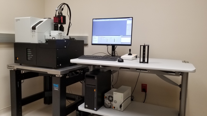
- 100-slide capacity for high throughput Imaging
- Transmitted light brightfield and epi-fluorescence scanning
- Automated sample identification and focus.
- Advanced stitching for tiled montage images
Fluorescence light source: LED Lumencor SOLA
Digital monochrome camera: Hamamatsu ORCA Flash4.0 V2
Color camera: 2/3” CCD camera, 3.45 μm x 3.45 μm pixel size
Fluorescence filters:
- DAPI ZERO LED: Ex392/23, Em447/60, Dichroic409 (DAPI, AF405, BFP, Pacific Blue)
- FITC ZERO LED: Ex474/27, Em525/45, Dichroic495 (FITC, GFP, Cy2, AF488)
- MCherry ZERO LED: Ex578/21, Em641/75, Dichroic596 (mCherry, TexasRed, mRFP)
- Cy5 ZERO LED: Ex635/18, Em680/42, Dichroic652 (Cy5, AF647, APC)
Objectives
| Objective | Mag. | NA | Immersion | WD (μm) |
|---|---|---|---|---|
| PlanApo N | 2x | 0.08 | dry | 6200 |
| UPlanSApo | 10x | 0.4 | dry | 3100 |
| UPlanSApo | 20x | 0.75 | dry | 600 |
| UPlanSApo | 40x | 0.95 | dry | 180 |
OLYMPUS FLUOVIEW FVMPE-RS Multiphoton LASER SCANNING MICROSCOPE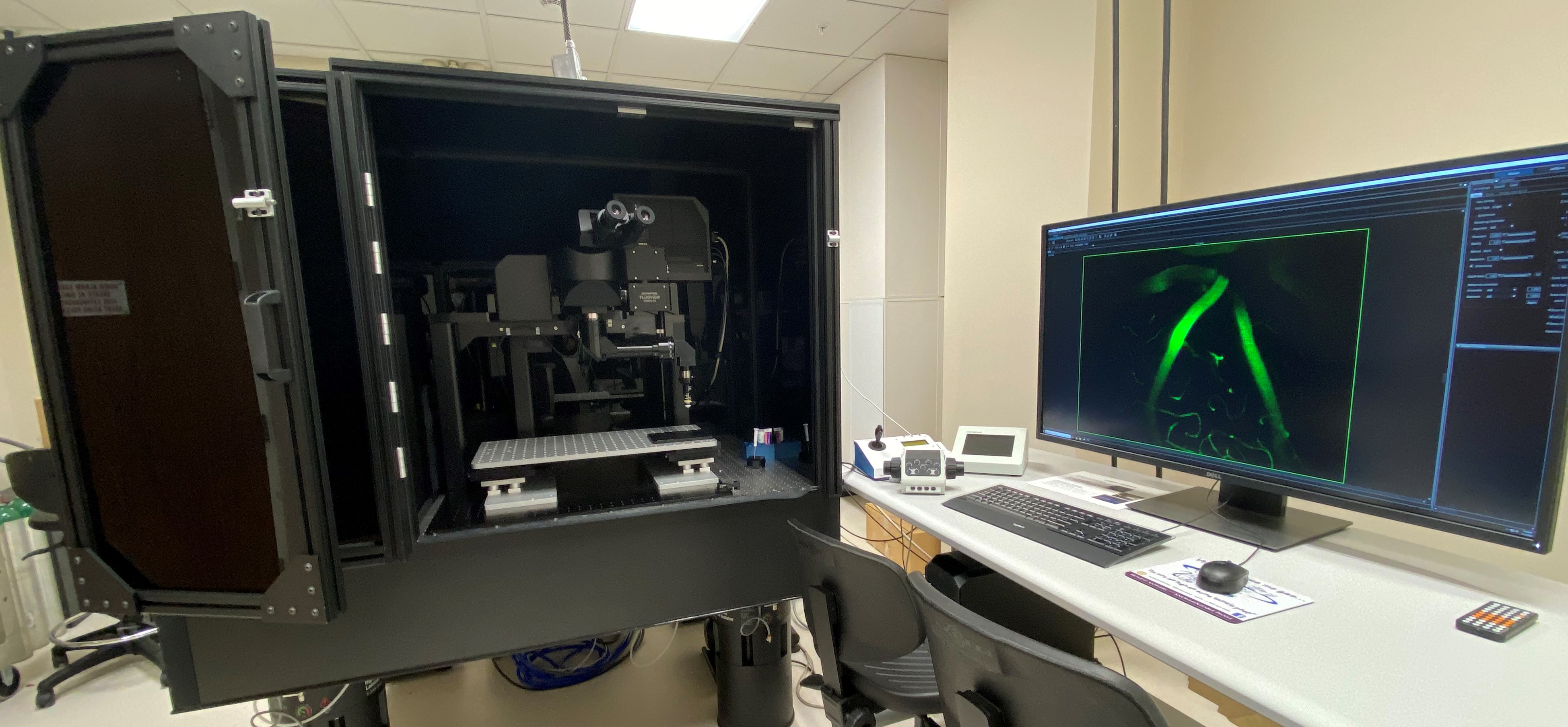
- Upright microscope with gantry frame, optimized for intravital imaging with a large space for live animals and experimental equipment
- Inner-focus articulating nosepiece will image at any angle
- Dual wavelength laser with a tunable range from 680 nm to 1300 nm and a fixed wavelength line at 1045 nm enables simultaneous multi-color, multiphoton imaging
- Resonant scanner for high-speed acquisition and galvonometer scanner for high definition
- 2 High-Sensitivity GaAsP detectors
Available Laser Lines (nm): Tunable 680-1300, Fixed 1045
Objectives
|
Objective |
Mag. |
NA |
Immersion |
WD (μm) |
|
UMPLFLN 10XW |
10X |
0.3 |
Water |
3500 |
|
XLPLN25XWMP2 |
25X |
1.05 |
Water |
2000 |
|
LUMPLFLN40XW |
40X |
0.8 |
Water |
3300 |
Filter cubes for eye viewing only
- DAPI 395/25X, BS425, 460/50M
- EGFP/FITC/CY2 470/40X, BS495, 525/50M
- DSRED/TRITC/Cy3 545/30X, BS570, 620/60M
Leica SP2 Confocal LASER Scanning Microscope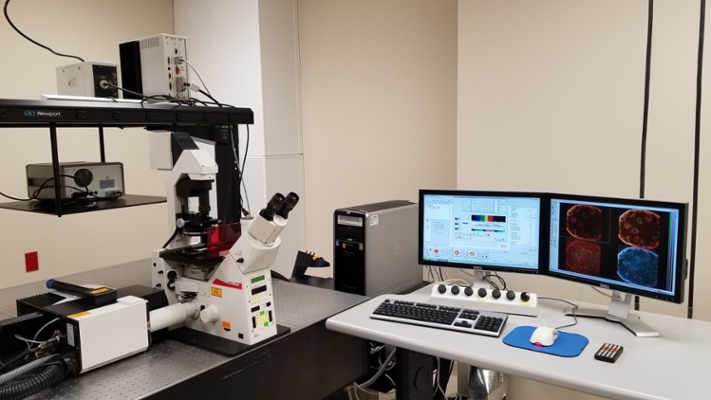
- Leica DMIRE-2 motorized inverted microscope
- AOBS spectral confocal scanning head
- R9624 Hamamatsu Photonics PMTs
Available Laser Lines (nm): 458-476-488-496-514-543-594-633
Objectives
| Objective | Mag. | NA | Immersion | UV | Working Distance (μm) | IC prism | Color ID |
|---|---|---|---|---|---|---|---|
| HC PL APO CS | 10x | 0.4 | dry | yes | 2200 | A | Yellow |
| HC PL APO CS | 20x | 0.7 | dry | yes | 590 | C | Green |
| HCX PL APO CS | 40x | 1.25 | oil | yes | 100 | E | Blue |
| HCX PL APO CS | 63x | 1.4 | oil | no | 100 | E | Dark blue |
| HCX PL APO CS | 63x | 1.2 | water | yes | 220 | - | Dark blue |
| U APO 340/cc | 40x | 1.15 | water | yes | 260 | - | Rubber cap |
Fluorescence filters for eye viewing only:
- I3 Cube: Ex BP450-490, Dichroic 510, Em LP 515 (FITC, GFP, Cy2, AF488)
- N2.3 Cube: Ex BP515-560, Dichroic 580, Em LP 590 (mCherry, TRITC, RFP)
NanoFluor II Integrated Microscope System
- IX-81 Olympus inverted microscope with focus stabilizer
- Yokogawa CSU-22 motorized confocal scanning head
- Ultra-fast spinning disk confocal with Z-piezo control (up to 100 frames per second)
- TIRFM (Total Internal Reflection Fluorescence Microscopy) attachment
- Atomic Force Bioscope SZ closed loop I
- Ar/Kr gas laser
Available LASER Lines (nm)
- Spinning Disk:488-514-568-647
- Ultra-fast Sinning Disk: 488-568
Objectives
| Objective | Mag. | NA | Immersion | UV | Working Distance (μm) | Color ID |
|---|---|---|---|---|---|---|
| U PL FLN | 10x | 0.3 | dry | no | 10000 | Yellow |
| U PL FLN PH | 40x | 0.75 | dry | no | 510 | Light blue |
| U PL FLN | 40x | 1.3 | oil | no | 200 | Light blue |
| U PL S APO | 100x | 1.4 | oil | no | 120 | white |
| PLAPON TIRFM | 60x | 1.45 | oil | no | 100 | Dark blue |
| PL APO | 60x | 1 | water | no | 150 | Dark blue & white |